MOON - in vitro endometriosis microphyisiological system
Ingénierie et Architecture
Endometriosis is chronic and debilitating condition that affects approximately 10% of reproductive aged women worldwide. It occurs when the endometrium, which lines the inside of the uterus, grows in areas outside the uterus, such as the peritoneum, ovaries and fallopian tubes. The misplaced tissue can cause severe pain, infertility and other morbidities.
The development of endometriosis is inevitably linked to menstruation, but its pathogenic mechanisms remain poorly understood. Historically, the most biologically relevant models of endometriosis are in menstruating non-human primates and in rodents. These carry significant costs and ethical concerns as well as limitations of translation to the clinic. Developing a more accurate and effective model to study endometriosis is essential to advance our understanding of the condition and establish better therapies.
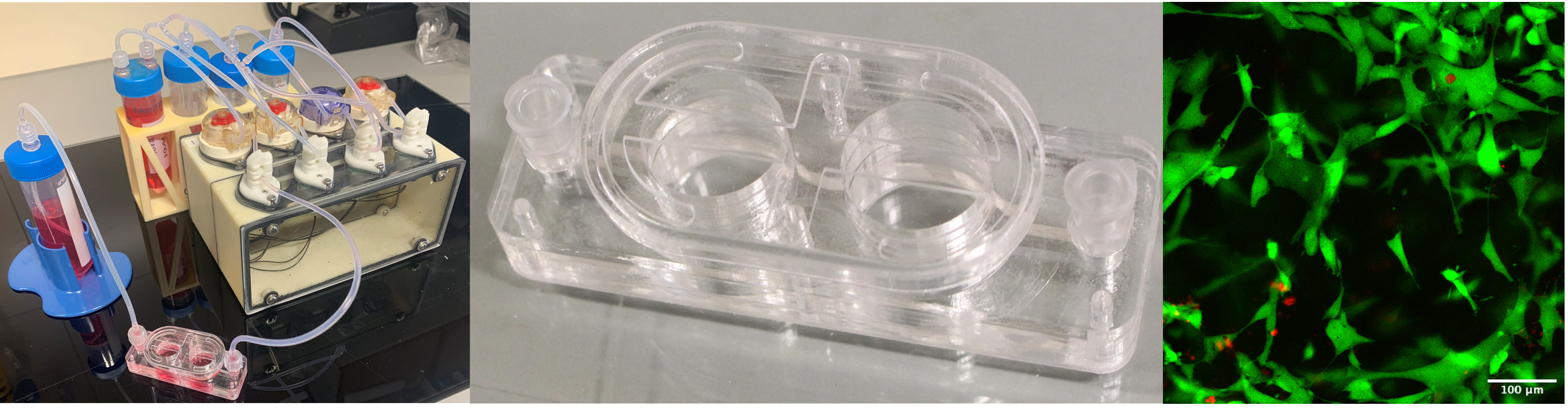
We aim to develop a sophisticated human-based in vitro microphysiological system (MPS) that accurately mimics the cyclical changes of the endometrium under the influence of hormones and answers the ethical, economical and scientific concerns associated with animal experimentation. Our MPS consists of three interconnected modules, each dedicated to simulating different aspects of the female reproductive system, with emphasis on endometriosis. The first module concerns a programmable pump system that delivers reproductive hormones at different concentrations according to each stage of the menstrual cycle to a second module, where a human-based 3D model of the endometrium is cultured. Models of typically affected tissue are cultured in the third module, where invasion by the endometrial tissue occurs. Altogether, the MPS is designed for microscopy imaging in a non-invasive manner, but it also allows tissue recovery for study of protein and gene expression.
We find that 3D co-culture models of human endometrium epithelial and stromal cell lines recapitulate features of human menstruation when exposed to different concentrations of estrogen and progesterone. Preliminary data suggests that fibroblast and intestinal cell constructs can be invaded by endometrial cells causing an increment of cell death. This showcases the feasibility of building a biologically relevant non-animal method for the study of endometriosis that can foster research of new drug targets and biomarkers in pharmaceutical, biotech and academic institutions.